How Secondary Antibody Selection Impacts Sensitivity in Neurodegenerative Disease Studies?
Neurons are known as the building blocks of the brain, spinal cord, and nerves. They act like communication units that control everything from thinking and memory to movement and sensation.
However, genetic mutations, abnormal protein build-up, mitochondrial dysfunction, oxidative stress, neuroinflammation, toxic environment exposure, traumatic brain injury, and age can gradually damage the neurons. As a result, the nerve cells lose their function and structure and eventually die.
Since neurons don’t regenerate easily, they can cause irreversible damage to the nervous system, leading to various neurodegenerative disorders.
These include:
- Alzheimer’s disease (AD)
- Parkinson’s disease (PD)
- Huntington’s disease (HD)
- Amyotrophic lateral sclerosis (ALS)
- Frontotemporal dementia (FTD)
Common symptoms of neurodegenerative disorders are:
- Memory loss or confusion
- Difficulty speaking or understanding
- Muscle stiffness, tremors, or weakness
- Loss of coordination or balance
- Mood and behavior changes
In order to treat these diseases, early diagnosis is important. Here is where different types of secondary antibodies come into play.
What Happens During a Neurodegenerative Disorder?
During neurodegenerative disorders, specific proteins start to misfold, clump together, and build up in the brain.
For instance, in:
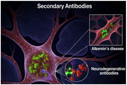
- Alzheimer’s disease: Amyloid-beta plaques form outside neurons, and tau protein tangles form inside neurons.
- Parkinson’s disease: Alpha-synuclein proteins clump together to form Lewy bodies inside neurons.
- Huntington’s disease: Mutant huntingtin protein accumulates and damages nerve cells.
- ALS: Abnormal aggregates of proteins like TDP-43 disrupt motor neurons.
These abnormal protein changes disrupt how neurons work and eventually kill them.
Researchers need to detect disease-specific proteins in order to treat a disease. This requires highly sensitive laboratory techniques like immunohistochemistry (IHC), western blotting, enzyme-linked immunosorbent assay (ELISA), and immunofluorescence (IF). All of these techniques rely heavily on secondary antibodies.
What are Secondary Antibodies?
Secondary antibodies are lab-made antibodies that attach to primary antibodies. The primary antibody binds directly to the target protein, while the secondary antibody binds to the Fc region of the primary antibody.
The secondary antibody is usually linked to a marker, such as an enzyme or a fluorescent dye. This marker produces a signal that shows scientists where and how much of the target protein is present.
For example, in an ELISA test for Alzheimer’s research, a goat anti-mouse IgG+HRP secondary antibody might be used. Here, “IgG” refers to the type of antibody it binds to, and “HRP” (horseradish peroxidase) is the enzyme that produces a color signal for detection.
However, in neurodegenerative disease, target molecules may be expressed at very low levels or present in small, localized regions of the brain. So, it is crucial to choose an antibody with high sensitivity.
What Factors Affect the Sensitivity of Secondary Antibodies in Neurodegenerative Disorders?
The choice of secondary antibody directly affects the signal-to-noise ratio (SNR)—a critical determinant of sensitivity. So, here are the factors that directly impact the sensitivity and accuracy of detection:
Affinity and Specificity
Affinity is the measure of how tightly the secondary antibody binds to the primary antibody. So, a high affinity means stronger binding and stronger signals.
Whereas, specificity can be defined as the ability to bind only to the intended antibody type and species. Naturally, you’d want high specificity to reduce background noise.
Cross-Reactivity Control
Cross-reactivity happens when a secondary antibody binds to other proteins instead of the target. This can create false signals.
In brain studies, where there are many similar proteins, the chances of cross-reactivity are high. So, it is recommended to choose cross-adsorbed secondary antibodies that eliminate the risk of cross-reactivity during the experiment.
Signal Amplification Strategy
Some secondary antibodies are designed to carry multiple markers. This is done to boost the signal without increasing background noise.
For example:
- Biotin-streptavidin systems amplify signals by attaching many biotin molecules to a single secondary antibody.
- Enzyme-linked systems like HRP or alkaline phosphatase can also amplify color or light output.
Since target proteins can be extremely rare in neurodegenerative disease, amplification is necessary for the research.
Choice of Detection Label
Each secondary antibody comes with an attached label. This helps determine how the signal is seen. For instance:
- Enzymes (HRP, alkaline phosphatase) produce a color or light signal.
- Fluorescent dyes (FITC, Alexa Fluor) emit light under specific wavelengths.
- Chemiluminescent labels emit light detected by special cameras.
In order to get reliable results, you need to choose the detection label carefully. And this depends on:
- The detection method (ELISA, IHC, IF, etc.)
- The required sensitivity level
- Whether multiple targets will be detected at once
Host Species Matching
Secondary antibodies must be raised against the same species as the primary antibody. It means if the primary antibody is from a mouse, you need an anti-mouse secondary antibody. Otherwise, this can to weak or no signal, which further reduces sensitivity.
Polyclonal V/S Monoclonal Secondary Antibodies
Polyclonal secondary antibodies bind to multiple sites on the primary antibody. This can increase signal strength.
In contrast, monoclonal secondary antibodies bind to one specific site. While this reduces the background signal, it may give a weaker signal.
So, in low-target situations, polyclonal antibodies may provide better sensitivity. However, monoclonal antibodies offer cleaner results when background noise is a concern. So, choose your secondary antibody while considering your preferences.
The Bottom Line
In neurodegenerative disease research, the amount of proteins linked to the disease can be very low. This makes it difficult to detect. However, choosing the right secondary antibody can help detect proteins even in low amounts. So, choose your secondary antibody while considering the aforementioned factors to get accurate and reliable results.